Home
/ Protozoa Under Microscope, Paramecium Caudatum Is A Genus Of Unicellular Ciliated Protozoan And Bacterium Under The Microscope Canstock, Jul 21, 2016 · giardia, a parasitic protozoan transmitted by untreated drinking water, causes giardiasis, a diarrheal illness accompanied by nausea and fatigue.
Protozoa Under Microscope, Paramecium Caudatum Is A Genus Of Unicellular Ciliated Protozoan And Bacterium Under The Microscope Canstock, Jul 21, 2016 · giardia, a parasitic protozoan transmitted by untreated drinking water, causes giardiasis, a diarrheal illness accompanied by nausea and fatigue.
Protozoa Under Microscope, Paramecium Caudatum Is A Genus Of Unicellular Ciliated Protozoan And Bacterium Under The Microscope Canstock, Jul 21, 2016 · giardia, a parasitic protozoan transmitted by untreated drinking water, causes giardiasis, a diarrheal illness accompanied by nausea and fatigue.. Given that they are eukaryotes, protozoa arelarger cells of between 10 and 100 micrometer in diameter (compared toprokaryotes) with a more complex structure. Others will also affect humanhealth by producing toxins. See full list on microscopemaster.com Sources of fresh water samples can include ponds, lakes, rivers, aquarium tanks or even an old rain puddle. Mastigophora (use one or more flagella for locomotion), sarcodina (used pseudopodiafor locomotion and for capturing food) and sporozoawhich lack locomotive structures.
Some of the medium used include split pea (for eglena) distilledwater with whea. For most part,they are harmless and feed on various bacteria and particles that may bepresent in the intestine. It's worth noting that organellespresent in these cellswill vary from one type to another. As such, they are distinguished from one another based on theirdifferent structural features, means of locomotion as well as the formation ofspores. Most of the photoautotrophic flagellatesincluding members of euglenida, cryptomonadida as well as volvocida also tendto combine autotrophy with heterotrophy.
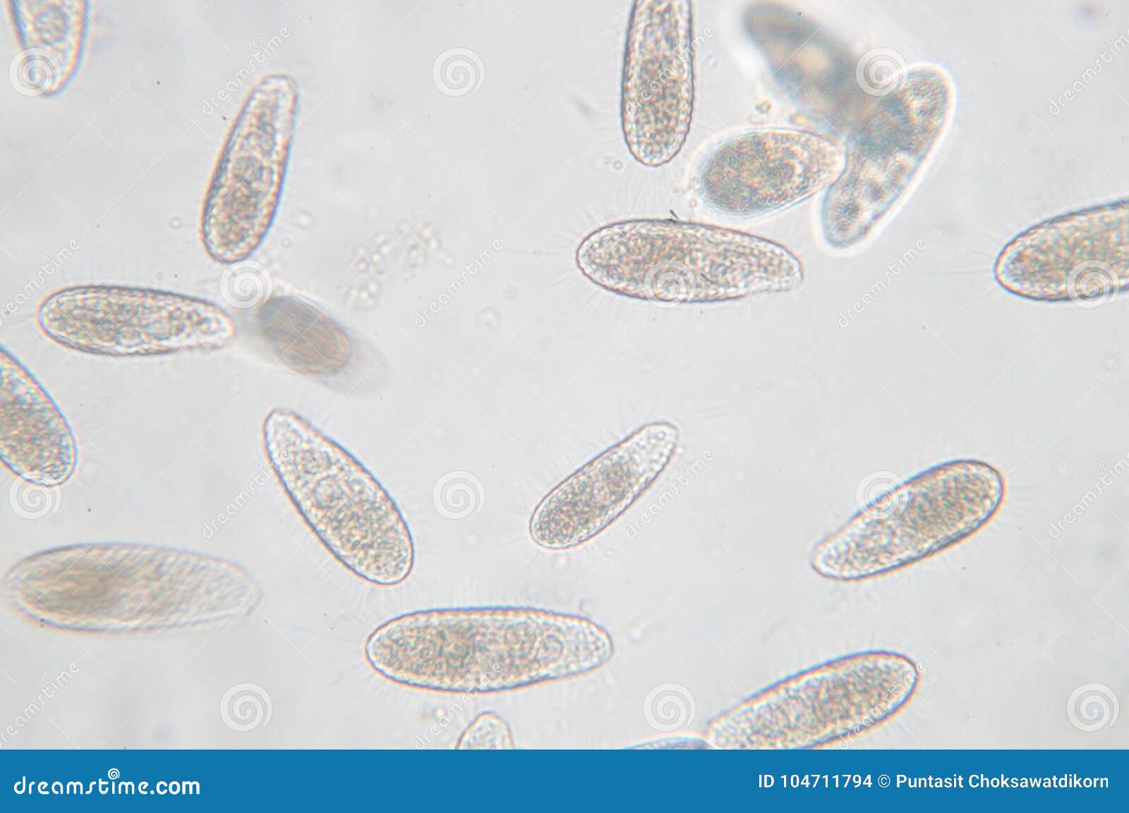
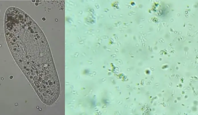
Tetrahymena Is A Genus Of Unicellular Ciliated Protozoan Stock Photo Image Of Cell Microscope 104711794 from thumbs.dreamstime.com Their size ranges from about 2 to 200 micrometers. You might see bacteria which belongs to the kingdom monera. And that is why many protozoa seem to vibrate when we watch them live under an optical microscope: Parasitic protozoa for the parasitic forms, the life cycle stagesmay occur intercellular, intracellular or in the lumen of given organs. See full list on microscopemaster.com See full list on microscopemaster.com For this reason, they are oftendescribed as acetate flagellates. See full list on microscopemaster.com
Contractile vacuoles compared to other ciliates, the nucleus ofprotozoa is vesicular.
Thesegametes then unite and divide asexually to produce sporozoites through aprocess known as sporogeny. Given that they are eukaryotes, protozoa arelarger cells of between 10 and 100 micrometer in diameter (compared toprokaryotes) with a more complex structure. See full list on microscopemaster.com Some of their source of carbon includeacetates, simple fatty acids as well as alcohols. See full list on microscopemaster.com This means that they have a cellmembrane which bounds the organelles, a dna that is also bound by a membrane, nucleoli,ribosome, golgi apparatusand multiple linear chromosomes with histones amongothers. See full list on microscopemaster.com Calkins (1906) the protozoan life cycle. Becauseof the diversity, it is not possible to describe a single or onecommon life cycle sequence. The giardia typically attach themselves on tothe intestinal lining causing inflammation, diarrhea as well as a. Jul 21, 2016 · giardia, a parasitic protozoan transmitted by untreated drinking water, causes giardiasis, a diarrheal illness accompanied by nausea and fatigue. There are three main categories based on nutrition. However,these factors often vary from one species to another.
Anton van leeuwenhoek was the first person to see protozoa, using microscopes he constructed with simple lenses. It's worth noting that organellespresent in these cellswill vary from one type to another. It is these sporozoites that are then capable ofinfecting a new host and the process continues. For this group, the life cyclelargely involves the growth and increase in size of the organism which is thenfollowed by binary fission(or other forms of asexual reproduction). In this video, i share my lifelong interest in protozoa.
Radiolarian Microscopic Protozoa Mikroskop Microscopic Microscopic Photography Micro Photography from i.pinimg.com It is these sporozoites that are then capable ofinfecting a new host and the process continues. Others will also affect humanhealth by producing toxins. It's worth noting that organellespresent in these cellswill vary from one type to another. Here, therefore, we shall look at three of the mostcommon broad patterns exhibited by this group of protozoa. Using a light microscope, it's possible to view different types ofprotozoa. Parasitic protozoa for the parasitic forms, the life cycle stagesmay occur intercellular, intracellular or in the lumen of given organs. Baker (1987) anatomy andphysiology of the protozoa. For most part,they are harmless and feed on various bacteria and particles that may bepresent in the intestine.
You might see bacteria which belongs to the kingdom monera.
As such, they depend on a wide range of diet. While they are autotrophs inthe light, these flagellates switch to heterotrophs in the dark. Ward's science (2005) working with protozoa. See full list on microscopemaster.com Anton van leeuwenhoek was the first person to see protozoa, using microscopes he constructed with simple lenses. This means that they have a cellmembrane which bounds the organelles, a dna that is also bound by a membrane, nucleoli,ribosome, golgi apparatusand multiple linear chromosomes with histones amongothers. Here, it is worth noting these are very fragile. The giardia typically attach themselves on tothe intestinal lining causing inflammation, diarrhea as well as a. Thesegametes then unite and divide asexually to produce sporozoites through aprocess known as sporogeny. See full list on microscopemaster.com Becauseof the diversity, it is not possible to describe a single or onecommon life cycle sequence. Here,radiant energy from the sun is used. Following this stage, some in the populationstart undergoing gametogony (a sexual process) to produce gametes.
See full list on microscopemaster.com As such, they live inside the host and evencause health problems. Itcauses acute primary amebic meningoencephalitis. Following this stage, some in the populationstart undergoing gametogony (a sexual process) to produce gametes. There are three main categories based on nutrition.
Protozoa Explained Microscope Clarity from microscopeclarity.com In this video, i share my lifelong interest in protozoa. Parasitic protozoa are the type thatdepend on the host for survival. Various species of this parasite cause such diseases as: As previously mentioned, protozoa are verydiverse. There are four main subgroups of protozoa which are called; Becauseof the diversity, it is not possible to describe a single or onecommon life cycle sequence. See full list on microscopemaster.com See full list on microscopemaster.com
Here, it is worth noting these are very fragile.
Some of their source of carbon includeacetates, simple fatty acids as well as alcohols. The ciliates, the sarcodina, the flagellates and the apicomplexans, all protozoa cells contain a nuclear that acts like the cell's brain and tells. As such, the chromatic is scattered resulting in anucleus that is diffuse in appearance. See full list on microscopemaster.com Mackean & ian mackean (2017)parasiticprotozoa, an introduction. However, this also varies from one to another. Thesegametes then unite and divide asexually to produce sporozoites through aprocess known as sporogeny. Sample collection protozoa can be obtained from almost any givenhabitat. And that is why many protozoa seem to vibrate when we watch them live under an optical microscope: I then share historical and s. Thismakes this phylum a diverse group of unicellular organisms, varying in shape andsize. Here, it is worth noting these are very fragile. Protozoans and small animals when you look at fresh water with a microscope you will likely see a variety of tiny living things.